The Mouse Brain In Its Colours Screensaver
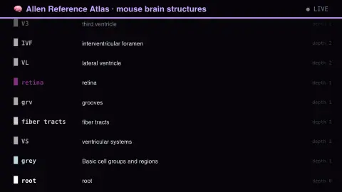
The The Mouse Brain In Its Colours screensaver shows structures from the Allen Reference Atlas of the mouse brain, key-less. Every region carries an assigned colour used consistently across every figure, slice and dataset the institute publishes, so the hex triplet is the region's identity rather than decoration, and each row is drawn in its own. Depth is the nesting level: depth 1 is a major division, depth 9 a specific cortical layer. It's free, runs in your browser, and ESC exits.
Live preview
Press ESC to leave fullscreen. Settings are saved in your browser only.
How the The Mouse Brain In Its Colours screensaver works
The saver reads the source described above and draws it on a canvas — no account, no tracking.
Structures from the Allen Reference Atlas of the mouse brain, key-less. Every region carries an assigned colour used consistently across every figure, slice and dataset the institute publishes, so the hex triplet is the region's identity rather than decoration, and each row is drawn in its own. Depth is the nesting level: depth 1 is a major division, depth 9 a specific cortical layer.
It refreshes on a schedule, so what you see stays current.
Colours can be changed in settings; nothing is stored beyond your own browser.
The Allen atlas and its colour-coded brain
The Allen Institute for Brain Science, a non-profit research institute in Seattle founded in 2003, built public reference resources for the mouse brain. The Allen Mouse Brain Reference Atlas is annotated on Nissl-stained sections from a single adult C57BL/6 mouse at postnatal day 56. A reference atlas is a map: it divides a standard brain into named regions so that experiments from different laboratories can be compared in the same coordinates.
The regions form a tree, from large divisions down through nested subdivisions to specific areas and layers. Each structure record has an identifier, a full name, an acronym, a parent structure, a depth in the tree and a colour stored as a hex triplet. The root of the tree, the whole brain, has depth 0. Related regions tend to share families of hues, which makes the hierarchy easier to see at a glance.
The mouse is a standard model organism in neuroscience, so the atlas is widely used, but it is a reference for a standard adult brain rather than any individual's. Other species, including humans, have their own atlases and naming schemes, and mapping between them takes careful comparative work.
A worked example
Structures from the Allen Reference Atlas of the mouse brain, key-less. Every region carries an assigned colour used consistently across every figure, slice and dataset the institute publishes, so the hex triplet is the region's identity rather than decoration, and each row is drawn in its own. Depth is the nesting level: depth 1 is a major division, depth 9 a specific cortical layer.
Settings & tips
- Accent and Background recolour the display.
- Every value on screen comes from the live source — nothing is simulated.
- Press ESC to leave fullscreen.
Frequently asked questions
- Is this real data?
- Yes — it comes from the source named on this page and is refreshed on a schedule.
- Is a key needed?
- No. This source is fully key-less.
- Is it free?
- Yes, and it runs entirely in your browser.
- Why use the mouse brain?
- Mice are a standard laboratory model, so a detailed reference map serves many research groups at once.
- What is the acronym next to each name?
- It is the atlas's short label for the structure, used where the full name would be too long. Every structure in the Allen ontology has one.
- Is this a map of every neuron?
- No. It is a map of named regions and their hierarchy, not of individual cells or connections, which are covered by other datasets.